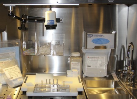
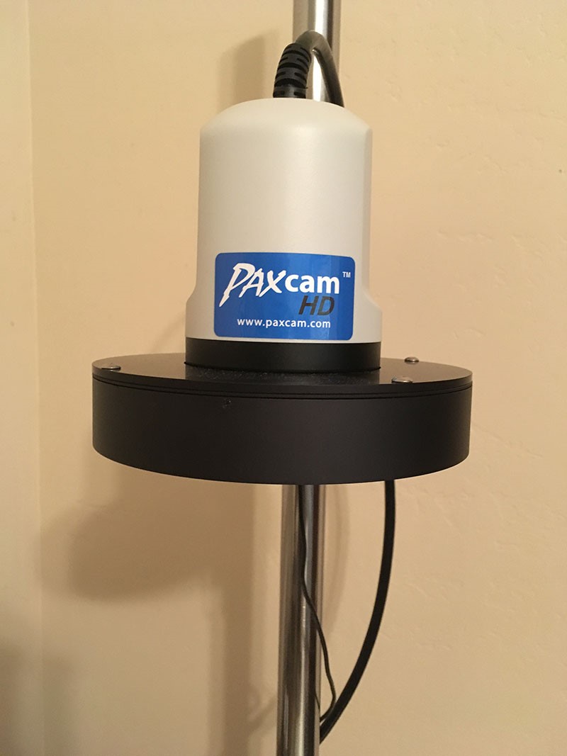
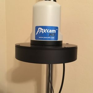
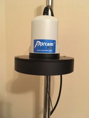
The PAXcam HD Grossing Station is designed to meet the specialized needs of the anatomic pathology grossing lab. The PAXcam Gross camera provides a stunning HD 1080p video view in real time, and the powerful 30x zoom range accommodates a full table view down to small fields of view within a tissue cassette. The zoom levels and other camera functions are controlled by footswitch for hands-free operation, using the ample presets provided. There is no need to touch the camera for any zooming, exposure, or focus controls.
Digital image capture of your surgical pathology specimens can be to external applications and/or to our PAX-it image management system. Video streams of live action may also be captured, and even streamed in real time for easy remote consultations on your network or over the internet! The flexibility of the system for image capture and working with live video allows for a host of pathology lab needs to be met with one easy-to-use system.
Marking up captured gross images is through simple touch controls, for measurements, sequential tagging of samples, and highlighting areas with circles, boxes, and text. Let us show you how easily these steps can be accomplished!
Image Analysis Software
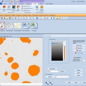
- The PAX-it Image AnalysisSoftware Module includes all the features of the Basic Measurement Module, while adding an additional level of capability. Rather than drawing measurements on an image, the image analysis tools automatically detect objects, layers, areas fractions, or optical profiles for data collection.
Flexible Routines for Photo Analysis
- PAX-it image analysis software is built flexible, allowing the user to define the specifics of the analysis in an understandable wizard format. No degree in computer science is required for these easy-to-use functions! Once analysis routines are defined, they may be saved as a stored routine, and applied to other images with the click of a button.
- Materials Science labs will benefit from specific routines included in the image analysis package, such as coating thickness detection, porosity analysis, nodularity analysis, ferrite-pearlite calculations, grainsizing, flake size distribution, and more. Specify any of these analyses according to your lab’s needs, and even design your own routine for area fractions or detection of objects via thresholding, to customize your data collection and reporting.
Automatically Analyze Your Images
- PAX-it! Image Analysis tools can use density, color, shape factors, and size filters to detect and sort objects or areas within your images. Filters may be applied to disregard objects of certain shapes or sizes, or to split the results into bins related to specific measurements.


