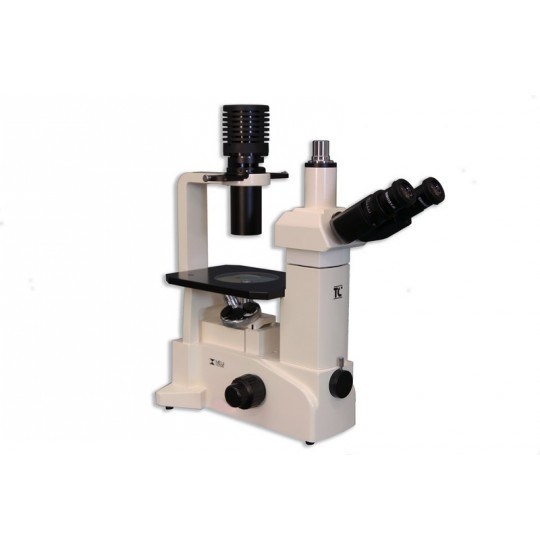
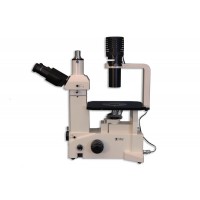
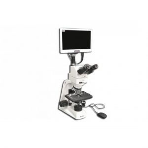
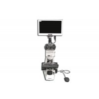
Features of the TC-5000 bright-field contrast microscope include:
The glasses are supplied by a standard Siedentopf observation binocular with a 30o angle or a tri-eyed observation head (connected to the camera) with a 45o angle
Equipped with 6V, 30W halogen lighting, 0.30N condensing device, KAK size 180mm (X) x 245mm (Y) with replaceable glass insert with 45mm opening, Integrated self voltage sensor power supply Dynamic power with local power cord
• Compact wedge-shaped aluminum alloy frames for exceptional stability and small footprint
• Chemical resistant enamel surface
• Flexible design and modularity for system expansion
• Transparent sample plate for easy target identification, Exchange observation tube, Automatic voltage sensor power supply with local power cord Low coaxial control and fine focus with adjustable levels 30W halogen lamp, CCD camera, analog or SLR mounted at the front
Bright field
This is the simplest of all optical microscope illumination techniques. Illuminate the transmitted sample (that is, illuminated from below and observed from above) white light and the contrast in the sample is caused by the absorption of some light transmitted in thick areas characteristics of the sample. Bright field microscopes are the simplest of the techniques used to illuminate patterns in light microscopy and its simplicity makes it a popular technique. Typical image of a bright field microscope image is a dark pattern on a light background.
Phase contrast:
This is an optical microscope technique that converts the light moving phases through a transparent specimen to change the brightness in the image. The displacement phases themselves are invisible, but become clear when displayed as brightness variations. When light waves travel through a medium other than a vacuum, the interaction with the environment causes the amplitude and phase of the waves to change in a way that depends on the properties of the environment. Changes in amplitude (luminance) resulting from scattering and absorbing light, are often dependent on wavelength and can give rise to color. Photographic equipment and the human eye are only sensitive to amplitude variations. There is no special arrangement, so the phase change is invisible. The TC-5300 & TC-5400 Series is suitable for viewing colorless and transparent specimens and living cells.
The TC-5300 & TC-5400 series is a contrast-enhanced optical technique that can be used to produce high-contrast images of transparent specimens such as living cells, microorganisms. , thin tissue slices, lithographic models and extra-cell particles (such as nuclei and other organelles). In fact, the phase-contrast technique uses an optical mechanism to convert minute-phase variations into corresponding amplitude changes, which can be visualized as differences in image contrast. One of the main advantages of phase contrast microscopes is that living cells can be examined in their natural state without being killed, fixed and stained. As a result, the dynamics of the biological processes taking place in living cells can be observed and recorded in high contrast to the clarity of minute sample details.


