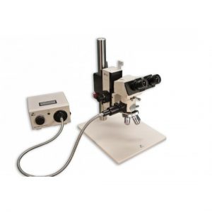
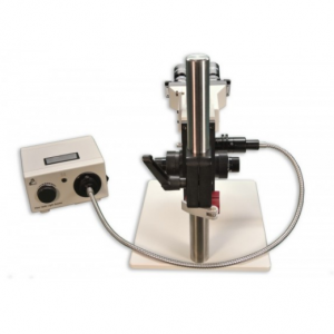
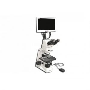
Description
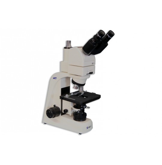
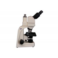
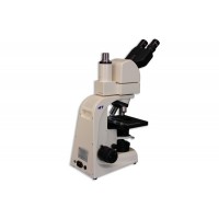
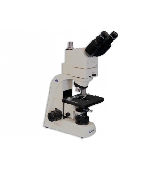
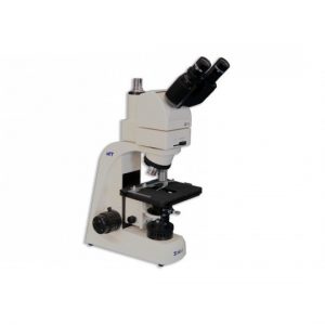
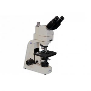
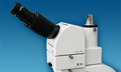
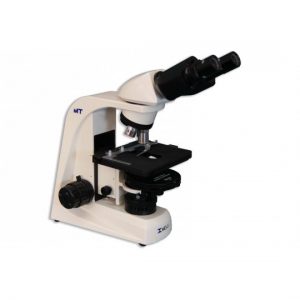
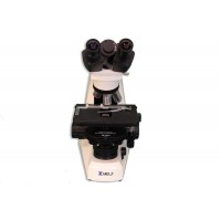
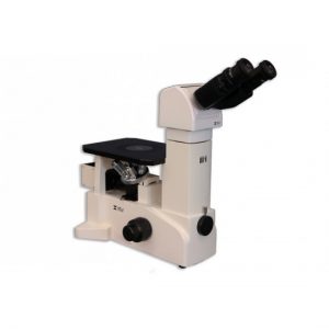
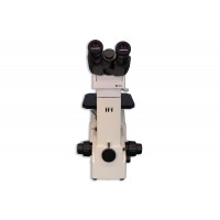
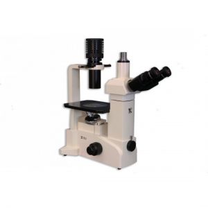
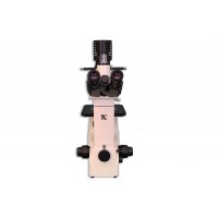
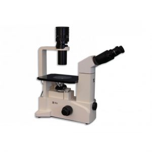
The MT4000 Microscopes features include:
- Compact Space Saving Footprint design with ergonomics in mind
- Infinity Corrected Optical System (ICOS™) with all newly designed Planachromatic optics
- Comfortable Siedentopf-type Binocular and Trinocular Viewing heads
- Optional Ergonomic tilting binocular head- Adjustable vertically from 10°to 50° degrees
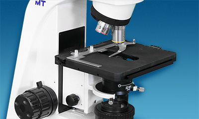
- Ceramic Coated Stage with Ergonomically Placed Low Position Drop down Control
- Parfocalled Microscope Objectives
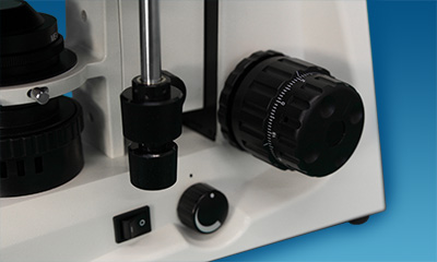
- Tension adjustment control with Coaxial knobs
- Low Positioned Ergonomic Coaxial Focus Control
- Low Positioned Ergonomic Coaxial Focus Control
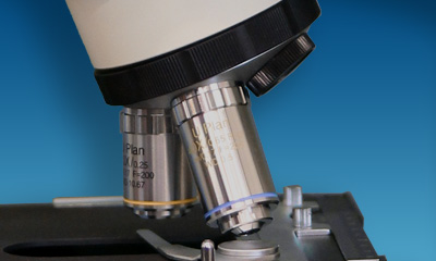
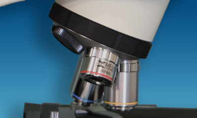
- Smooth Operating Ergonomic Quintuple Nosepiece
- Brightfield and Phase Contrast Observation modes available
- Super Bright LED Koehler illumination, lambertian design
- Integrated 30-Watt Halogen Transmitted illumination System on selected Brightfield and Phase Contrast Models
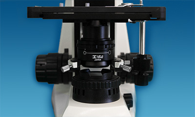
- Abbe and Zernike Phase Contrast Condensers available
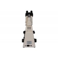
- Integrated Automatic Voltage Sensing Power Supply with localized power cord
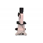
- Rear Cord Wrap for easy storage
- Wide Range of Filters and Accessories
Brightfield:
The simplest of all the optical microscopy illumination techniques. Sample illumination is transmitted (i.e., illuminated from below and observed from above) white light and contrast in the sample is caused by absorbance of some of the transmitted light in dense areas of the sample. Brightfield microscopy is the simplest of a range of techniques used for illumination of samples in light microscopes and its simplicity makes it a popular technique. The typical appearance of a brightfield microscopy image is a dark sample on a bright background.
The MT4000 Series is a newly designed upright biological compound microscope system. Ergonomically designed, precisely manufactured and simple to use stage and coarse and fine controls. To meet the demands of researchers and clinical laboratory specialists. The MT4000 Series was to develop to provide functionality and operational ease. Viewing high quality images, sample changing and photomicroscopy are all conducted with ergonomics in mind. High luminescent LED illumination reduces need for frequent bulb replacement but still provide rugged durability and long life span.
VIEWING HEADS
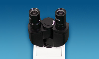
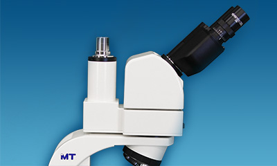
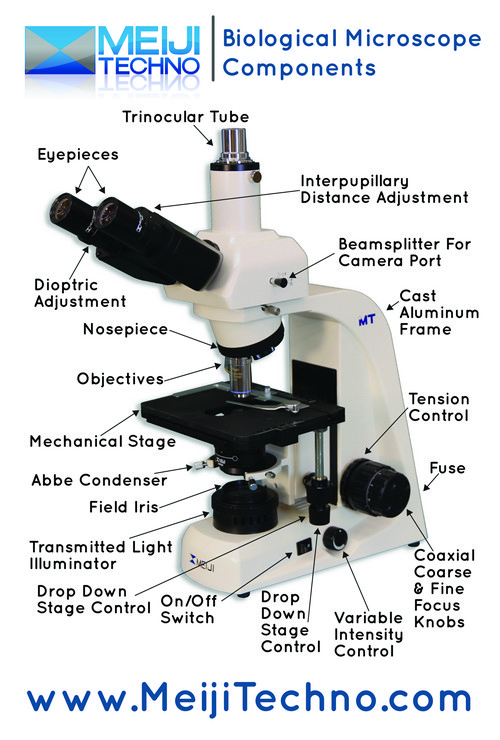
Model MA815/05 is the Siedentopf-type binocular head and Model MA816/05 is the trinocular head for camera integration. Each head has the eyetubes inclined at 30 ° with the left eyetube having graduated diopter settings. The interpupillary distance is adjustable between 53mm – 75mm. An 80/20 beamsplitter for the trinocular tube can be engaged for photo work.The optional MA957/05, Ergonomic Binocular Viewing Head, has the inclination adjustable from 10 to 50 degrees to fit users of different heights. The Ergonomic Trinocular model comes with the MA958, photo/video attachment with sliding 80/20 beam-splitter (Included in MT4300ED).
EYEPIECES
10X Widefield High Eyepoint eyepieces F.N.20 are standard, and 15X and 20X eyepieces are available as an option. A Widefield High Eyepoint 10X focusable eyepiece that accepts 21mm reticules is also available.
OBJECTIVE CHANGER
Quintuple nosepiece provides effortless objective changes with smooth-operating, ball bearing mounted.
OBJECTIVES
Meiji Techno America offers an assortment of Planachromat Infinity Corrected (ICOS) objectives for Brightfield, Darkfield and Phase Contrast observation modes.
STAGE
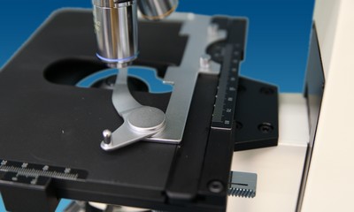
Ceramic coated standard right-handed or optional left-handed controls, flat top stage 171mm x 115mm and travels 78mm(X) x 52mm(Y). Ergonomically positioned coaxial drop down controls. Available with motorized focus and stage controls.
CONDENSER
New design centerable Abbe 1.25 N.A. Condenser with built-in iris diaphragm in dovetail mount.
ILLUMINATION
Powerful white LED illumination provides enhanced image quality and brightness for the observation of specimens and photomicroscopy.
PHOTOMICROGRAPHY
Meiji Techno offers a broad range of digital video cameras which offers excellent sensitivity, high resolution and a wide dynamic range for the most demanding brightfield and darkfield microscopy applications including clinical pathology and cytology, life sciences and geology. Some models has included software, that can preview and capture still shots and live videos to a PC or Mac with USB port or HDMI connectivity.


